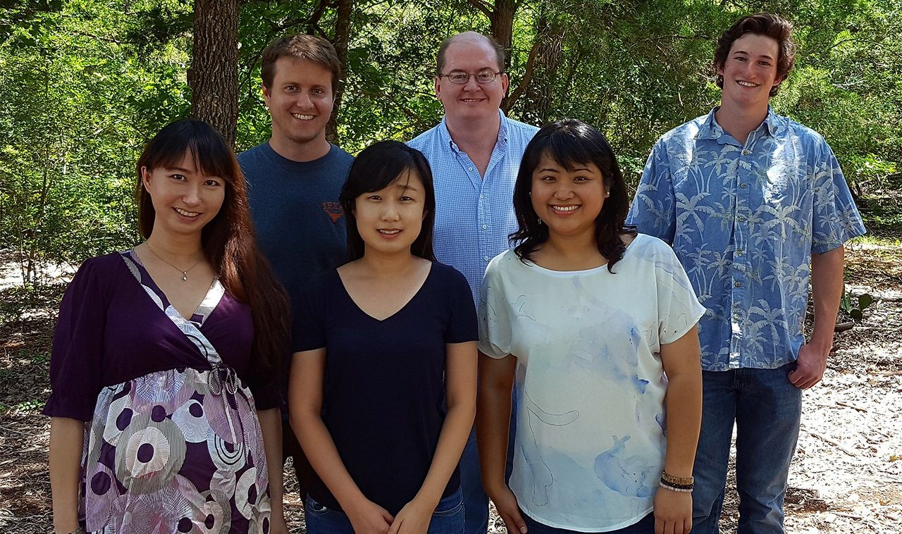
Life in the Lab
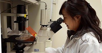
Inverted microscopes equipped with a camera, like the one Dr. Sunhee Lee is using, are ideal for observing and digitally imaging cells contained in tissue culture dishes.
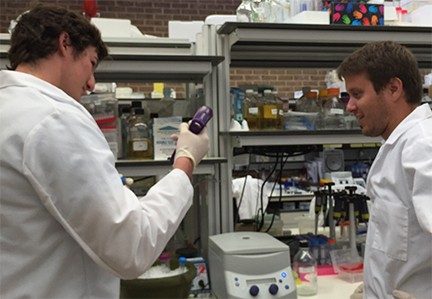
Postdoctoral fellow Daric Wible chats with summer intern Sebastian Bedford as Bedford harvests cultured tumor cells. Daric won the first place AMGEN Award in Basic Science Research for his platform presentation during MD Anderson Trainee Research Day in May, 2017. He presented that work at the Advances in Oncology Institutional Grand Rounds in June, 2017. In September 2019, he won a 2nd place poster award at the Texas Medical Center 9th Annual Postdoctoral Science Symposium, and 1st place at the Center for Molecular Carcinogenesis and Toxicology Research Symposium at UT Austin.
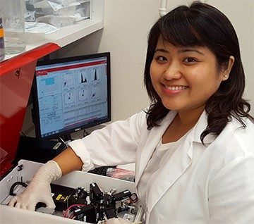
Former student, now postdoc, Sophie Chen, Ph.D., working with a flow cytometer. Flow cytometric methods provide a quick way to characterize different sets of cells in a larger population of cells. The Bratton lab uses flow cytometry to identify apoptotic cells.
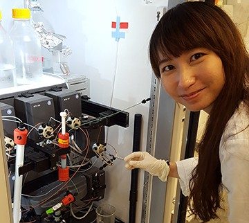
Dr. Crystal Wu is working with a fast protein liquid chromatography (FPLC) unit. Columns can be packed with different resins that will separate proteins based on characteristics such as size (gel filtration), charge (ion exchange) and affinity.