request an appointment online.
- Diagnosis & Treatment
- Cancer Types
- Lung Cancer
- Lung Cancer Diagnosis
Get details about our clinical trials that are currently enrolling patients.
View Clinical TrialsLung Cancer Diagnosis
Early stage lung cancer often does not have symptoms. In addition, when symptoms appear they can easily be mistaken for common respiratory illnesses like bronchitis or pneumonia. Because of this, many cases are diagnosed at an advanced stage.
Patients at high risk for lung cancer, especially those with a history of smoking, should undergo regular screenings in order to catch the disease at its early stages, when there is a better chance of cure.
If you have symptoms that signal lung cancer, your doctor will ask you questions about your medical, smoking and family history and whether you have been around certain chemicals or substances.
You will then undergo an imaging exam, typically a chest X-ray. Images cannot diagnose lung cancer, but they can show areas of concern. If the image shows such an area, the doctor may order other scans, including a CT scan or PET scan, for additional details regarding the area of concern.
If the findings on the imaging scans indicate cancer, the doctor will request that tissue or fluid be removed from the lung for examination. The act of obtaining a tissue or fluid sample is called a biopsy. There are several ways doctors can perform biopsies of lung tumors:
- Needle biopsy: A CT-guided biopsy where a needle is inserted through the skin under local anesthesia to acquire a tumor sample. One type of needle biopsy is fine needle aspiration (FNA), which uses a very small needle and suction to remove a small amount of tissue.
- Thoracentesis: Fluid from around the lungs is drawn out with a needle and tested for cancer cells.
- Bronchoscopy: A thin, flexible tube with a tiny camera is inserted through the nose or mouth and down into the lungs to obtain a small tissue sample (biopsy). This is usually performed under mild sedation. Bronchoscopies are rarely done alone. Bronchoscopy is usually performed with an endobronchial ultrasound.
- Endobronchial ultrasound (EBUS): A bronchoscope with an attached ultrasound device is used to check for lung cancer inside nearby chest lymph nodes. EBUS is often performed at the same time as a bronchoscopy and requires general anesthesia.
- Video-assisted thoracoscopic surgery (VATS): This minimally invasive surgical procedure uses a small camera to help retrieve tumor samples that are otherwise difficult to access. VATS requires a general anesthetic and is performed in the operating room by a thoracic surgeon.
- Thorascopy/pleuroscopy: A thin, flexible tube with a tiny camera is inserted through a small incision in the back (for a thorascopy) or between the ribs (for a pleuroscopy). Doctors use this device to look for and retrieve suspected cancer tissue.
To complete assessment of how advanced the cancer is, which is called staging, the patient will undergo a PET-CT scan and an MRI or CT scan to check for signs of cancer spread to other organs, including the brain. This will guide the treatment decisions for each patient’s lung cancer.
In some cases, lung cancer can be passed down from one generation to the next. Genetic counseling may be right for you. Visit our family history site to learn more about genetic counseling and testing.

Bronchoscopy 101: How it helps diagnose and treat lung conditions
A bronchoscopy is a minimally invasive medical procedure in which doctors use a special scope to examine the inside of your lungs and airways. It is used frequently to diagnose and stage lung cancer.
But can a bronchoscopy tell us anything else? Do you have to be put to sleep to have one? And how long does it take to recover?
Read on, for answers to these questions and more.
What are the different types of bronchoscopy?
Diagnostic bronchoscopy
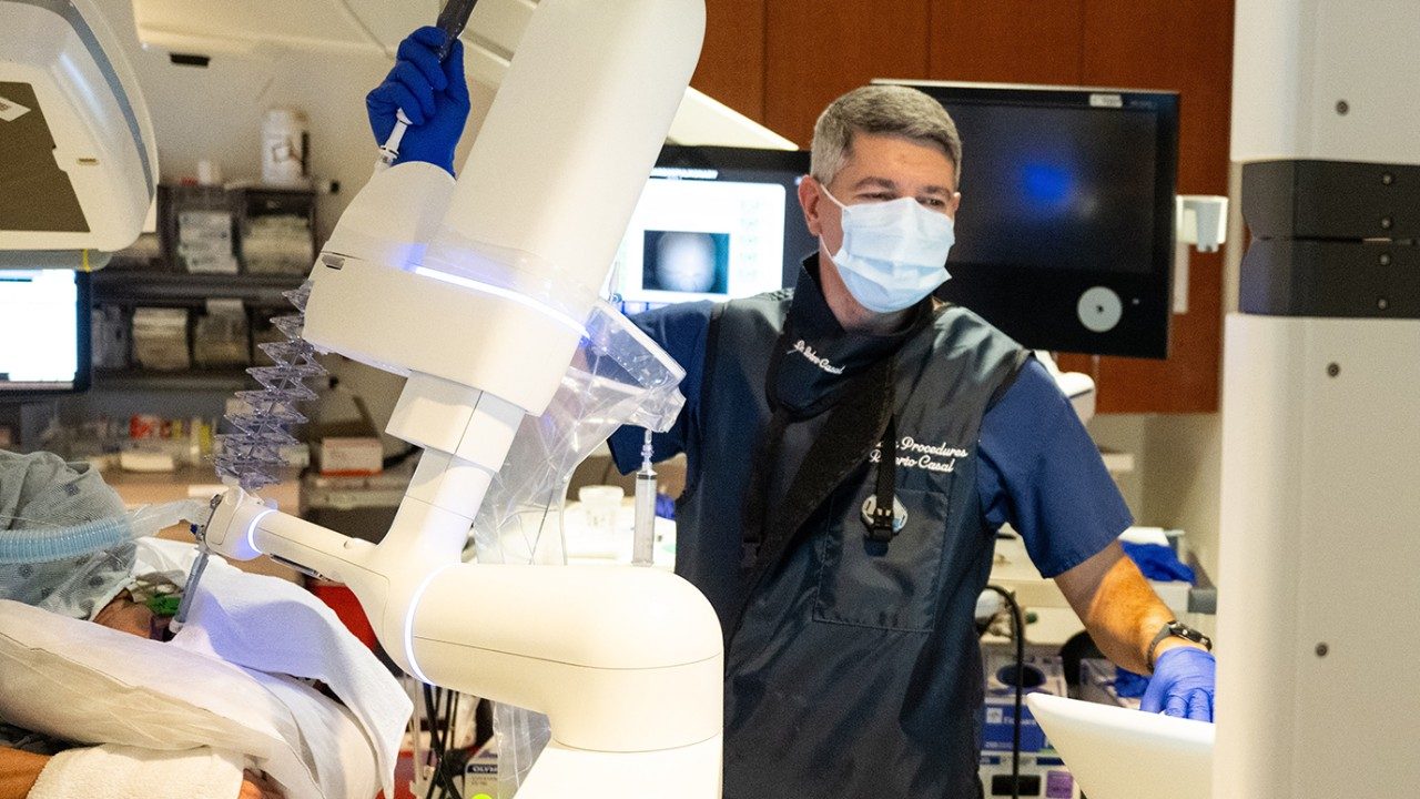
We use robotic bronchoscopy to biopsy lung nodules and a technique called endobronchial ultrasound (EBUS) to sample lymph nodes for cancer staging.
We also use a technique called bronchoalveolar lavage (BAL), typically in immunocompromised patients, to diagnose opportunistic infections of the lungs. To do a bronchoalveolar lavage, we wedge the bronchoscope in the section of the lung we’re interested in, flush it with a saline solution, extract the fluid, and culture it in a lab to see what grows.
Therapeutic bronchoscopy
Mostly performed via rigid bronchoscopy, this procedure is used to remove tumors that are blocking the windpipe, to cauterize bleeding tumors, or to place stents that keep the windpipe open. It won’t cure cancer, but it should make breathing easier.
How long does a bronchoscopy take?
That depends. The shortest procedure is the bronchoalveolar lavage, which can take just 10 to 20 minutes. The diagnostic bronchoscopy of a lung nodule can take 45 minutes to an hour, and the sampling of lymph nodes for staging can add another 45 minutes.
But if we’re doing it strictly for therapeutic purposes, it can take from one to two hours, depending on the complexity of the case.
Keep in mind, though, that these are all estimates. The length of each procedure is determined by the number of lung nodules or lymph nodes being biopsied, and doctors don’t know this until they are looking inside your lungs.
Are you awake during a bronchoscopy?
Not necessarily. You’ll be given general anesthesia in most cases, but moderate sedation if you only need bronchoalveolar lavage.
Is a bronchoscopy considered a serious procedure?
That depends on how you define it. I consider anything requiring general anesthesia to be a serious procedure.
But doctors will be working inside your lungs, which are the most vital organs aside from the heart. So, it is more serious than a colonoscopy, for example, but less serious than most surgeries.
Is a bronchoscopy considered a high-risk procedure?
Bronchoscopy for the diagnosis of lung nodules, sampling of lymph nodes, or bronchoalveolar lavage is commonly considered a moderate-risk procedure. On the other hand, therapeutic bronchoscopy is always considered high-risk because patients are sicker to begin with. They often have collapsed or obstructed airways, low oxygen levels, or they are actively bleeding.
What are the risks of a bronchoscopy?
With diagnostic and staging bronchoscopies, the risks are mainly from the anesthesia, rather than the procedure itself. General anesthesia can lower your blood pressure, which can be risky if you have underlying cardiovascular disease. It can also weaken the breathing muscles, so you tend to take shallower breaths when you wake up from anesthesia, and that can make your blood oxygen levels drop.
Biopsies of the lung nodules or lymph nodes have minimal risk of bleeding and infection, but biopsy of lung nodules can sometimes lead to lung collapse (pneumothorax). That’s why even if everything goes well, we prefer our patients to avoid long-distance traveling or air travel until the next day.
With therapeutic bronchoscopies, the risks are higher, and they generally involve bleeding, having low oxygen levels, and difficulty breathing. But all of these are dependent on each patient’s particular situation, and your doctor will go over which ones apply to you in detail.
How long does it take to recover from a bronchoscopy?
Most diagnostic and staging bronchoscopies are outpatient procedures. Patients remain in the recovery area for 45 minutes to an hour afterward, and then they go home. They generally feel tired on the day of their bronchoscopy, but they are back to their baseline the following day.
The recovery time is similar for therapeutic bronchoscopy patients, even if they are already admitted to the hospital.
Is a bronchoscopy painful?
No. The lungs have no pain receptors, so they do not hurt, and you are also sedated. You should not experience chest pain after bronchoscopy, either, unless you are coughing really hard.
Does a bronchoscopy have any side effects?
Some patients report having a sore throat due to the breathing tube. And patients can expect to cough up mucus tinged with blood for the next 2 or 3 days. But those are both normal and will resolve on their own.
Is a bronchoscopy better than a CT scan?
These two tests play very different roles. A CT scan is the test that generally finds out there is something wrong in your lungs. But it can only tell you that there’s an abnormality present. It cannot tell you exactly what it is. A CT scan is just the beginning.
For an accurate diagnosis, we have to examine tissue under a microscope. A bronchoscopy allows us to obtain that sample and make a diagnosis. If it turns out to be lung cancer, the tissue we obtain through a bronchoscopy can also be used for molecular profiling, so we can tailor your treatment to any specific genetic mutations. And bronchoscopy can, in addition, sample the lymph nodes to determine the stage of lung cancer.
Can a bronchoscopy detect tuberculosis?
Yes. Bronchoalveolar lavage is considered the gold standard for detecting any type of lung infection. For patients with pneumonia, we can determine exactly which pathogen is causing it.
What’s the one thing people should know about bronchoscopies?
Go to a place like MD Anderson which does a large number of complex bronchoscopies each year and has a lot of expertise in them. That level of experience will give you a better outcome.
MD Anderson is also at the forefront of using ablation therapy during bronchoscopy to treat small, peripheral lung cancers through clinical trials. We are the pioneers in this field, and only a handful of cancer centers offer it right now.
Roberto Casal, M.D., is an interventional pulmonologist specializing in bronchoscopies.
Request an appointment at MD Anderson online or call 1-877-632-6789.
Clinical Trials
MD Anderson patients have access to clinical trials
offering promising new treatments that cannot be found anywhere else.
Becoming Our Patient
Get information on patient appointments, insurance and billing, and directions to and around MD Anderson.
Help #EndCancer
Give Now
Donate Blood
Our patients depend on blood and platelet donations.
Shop MD Anderson
Show your support for our mission through branded merchandise.
