Instrumentation
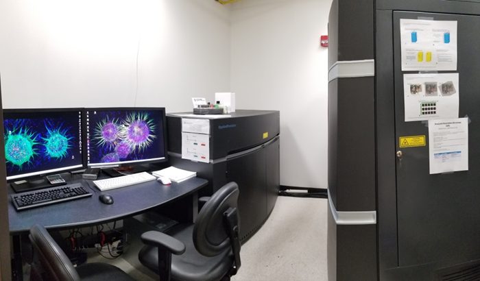
DeltaVision OMX Blaze V4 Super-Resolution Microscope

The OMX Structured Illumination Microscope (3D-SIM) provides super-resolution imaging for thin fluorescent slides (less than ~25 μm) and Total Internal Reflection Fluorescence (TIRF) imaging for live cells. It utilizes patterned illumination to extract high frequency information from fixed samples ~2x below the conventional resolution limit in 3-dimensions. RING TIRF provides selective illumination proximal to the coverglass (<100 nm) for high contrast/sensitivity detection of fluorescent molecule dynamics at the cell surface in live cell studies such as vesicle trafficking (exocytosis/endocytosis), receptor-ligand interactions, cellular adhesion and other membrane-related events. This microscope provides temperature control for live cells.
- Modes of operation:
- Structured Illumination (SIM)
- Total Internal Reflection Fluorescence (RING TIRF)
- Deconvolution
- Widefield
- Spatial resolution: ~120 nm XY and ~250 nm Z
- Optical Configuration “BGR”: 405 nm, 488 nm, 568 nm, 642 nm
- Best for: 3-D super-resolution imaging of fixed fluorescence-labeled cells and subcellular structures, 3-D deconvolution imaging of live cells, cellular adhesion, endo and exocytosis
Leica TCS SP8 Confocal Microscope with Environmental Chamber

Laser scanning confocal microscope for optical sectioning, z-stacking and tile scanning. Suitable for fixed and live cells, medium thickness tissue sections and organoids up to ~100 μm thickness. Unique advantages of this microscope include the fast resonant scanning option and interline scanning mode for dynamic/live events and colocalization, as well as flexible spectral detection for fluorescence fingerprinting, unmixing and for unconventional fluorophores. This microscope provides full environmental control with CO2 and humidity for live cells.
- Modes of operation:
- Conventional, point-by-point laser scanning
- Fast resonant scanning for living, motile cells
- Fluorescence Recovery after Photobleaching (FRAP)
- Automated mosaic stitching
- Reflection mode
- Spatial resolution: ~ 250 nm XY and 600 nm Z
- Available laser for fluorescence excitation: 405, 442, 458, 488, 514, 561, 633 nm
- Spectral detection with Acusto-Optical Beam Splitter (AOBS)
- Sensitive Hybrid Detectors (HyD)
- Inverted configuration
- Best for routine imaging of fixed cells and tissues; ideal for live cell confocal imaging due to low dwell reducing phototoxicity and maintaining cell viability.
Zeiss LSM880 Confocal Microscope with Airyscan

Laser scanning confocal microscope for optical sectioning, z-stacking and tile scanning. Suitable for fixed and live cells, tissue sections and organoids up to ~100 μm thickness. Unique advantages of this microscope include the Airyscan 32-hexagonal array detector enabling super-resolution. Each member of the array mimics a pinhole with an aperture of 0.2 Airy Unit, together collecting the entire point spread function, maximizing resolution and sensitivity.
- Modes of operation:
- Conventional, point-by-point laser scanning
- Airyscan array super resolution imaging
- UVA laser microirradiation
- 96 well plate scanning
- Photomanipulations (FRET/FRAP)
- Spatial resolution ~140 nm XY and ~400 nm Z
- Excitation lasers: 405, 488, 561, 594 and 633 nm
- 355 nm OPSL laser for DNA damage repair studies
- 32-channel GaAsP spectral detector (Airyscan) plus 2 PMTs and transmitted light PMT
- DIC/Nomarski optics to enhance transmitted light contrast
- XL incubator chamber and CO2 control for live cell and tissue imaging
- Best for high-resolution imaging of cells and medium thickness fluorescent specimens across the complete spectral range, and UVA laser microirradiations to assess live cell response to DNA damage.
Leica SP8 DIVE Multiphoton (2-Photon) Microscope

This microscope is preferred for thick tissues and intravital imaging. It enables deep imaging (up to ~500 μm) using infrared long wavelengths and highly focused z-spot illumination for 2- and 3-photon excitation and non-descanned detectors for efficient detection. Compared to the visible light, the infrared light penetrates deeper into tissues with increased imaging depth, minimal light scattering and reduced phototoxicity. The 2- and 3-photon effect provides high z-resolution without needing a confocal pinhole.
- Modes of operation
- Conventional galvo point scanning for highest resolution
- 8 kHz resonant scanning for highest speed (28 fps at 512 x 512)
- Non-linear optical imaging by second and third harmonics (SHG/THG)
- SpectraPhysics Insight X3 dual near infrared laser: tunable line from 680 nm to 1300 nm + 1040 nm fixed line for excitation of a range of fluorophores from blue to far red
- Variable dichroic-based spectral detection provides for simultaneous multi-channel acquisition
- 4 high QE non-descanned hybrid (HyD) detectors
- Heated animal platform and EZ-9000 anesthesia & nose cones for intravital imaging
- Objectives:
- 25X water immersion dipping objective (NA 0.95, w.d. 2.6 mm)
- 40X water coverslip corrected objective (NA 1.1, w.d. 0.65 mm)
- Upright configuration
- Scientifica stage and incubation with CO2 control
- Best for intravital imaging of dynamic in vivo processes and 3D (and 4D) volumetric imaging of thick tissue slices and organoids.
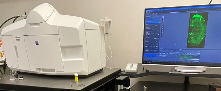
Zeiss Gaussian Lightsheet 7 (LS7) Microscope

This microscope is preferred for 3-D imaging of thick specimens such as biopsies or mouse whole organs (up to 20 mm) that are optically clarified using special tissue clearing procedures. Illumination orthogonal to detection optics generates a thin light sheet for selective planar illumination of the imaging plane, sparing the remainder of sample excitation thereby permitting rapid FOV camera-based imaging of the entire volume and minimizing sample bleaching.
- Modes of operation
- Dual sided lightsheet illumination
- Single sided lightsheet illumination
- 6 laser lines (405, 445, 488, 514, 561 and 638 nm) for rapid multispectral imaging from blue to far-red
- 5X and 10X illumination optics
- Detection optics include 5X and 20X water and clearing (mixed immersion)
- 2 high speed (57 fps at 1K x 1K) and high quantum efficiency (83% QE) PCO Edge 4.2 sCMOS cameras
- Best for rapid volumetric imaging of fixed and cleared whole organs, tissues and embryos.
LifeCanvas SmartSPIM Lightsheet Microscope

This microscope is preferred for mesoscale 3-D imaging of very thick optically clarified tissue specimens such as tumors or mouse/rat whole organs up to 40 mm. Illumination optics generate a thin light sheet for selective planar illumination microscopy (SPIM) that is axially swept across the sample for higher z-resolution and minimal vignetting and permitting rapid FOV camera-based imaging of the entire volume.
- Mode of operation: Dual illumination SmartSPIM using custom optics to axially sweep the light sheet beam waist across the sample for high-speed imaging of the sample volume
- 4 laser lines (445, 488, 561 and 638 nm) for rapid multispectral imaging from green to far-red
- 3.6X (N.A. 0.2 and w.d. 12 mm), 1.625X (N.A. 0.12 and w.d. 13 mm) and 9X detection optics
- Extended range sample carrier permitting sample size to 40 mm x 65 mm laterally.
- High QE fusion sCMOS camera
- Best for true meso-scale rapid volumetric imaging of fixed and cleared whole organs, tissues and embryos.
IN Cell Analyzer 6500 HS Slide/Plate Scanner

The IN Cell 6500 HS is a immunofluorescence slide and plate scanner for rapid high-throughput epifluorescence microscopic imaging.
- 4X, 10X, 20X, and 40X objectives
- Filter combinations for DAPI, AF488/GFP, AF568/RFP and AF647/Topro
- Requires #1.5 coverslip bottom plates (96-well, 48-well, 24-well, 12-well)
- Tile/z-stack capable
- Microfluidics (e.g., drug delivery) and equipped with incubation and CO2 control for live cell.
IMARIS 4-D Image Analysis and Tracking Software

- Volume Rendering
- Morphometric analysis
- Iso-Surfaces, Segmentation
- Feature detection (spots, cells, nuclei, organelles, foci, cytoplasm, cell membranes, neuronal dendrites, etc.)
- Object detection and 4D spatiotemporal tracking
- Colocalization analysis
- Image mathematics
- 3D/4D video design
Zeiss Axio Observer Z1

Epifluorescence microscope for capturing wide-field fluorescence images of fixed and live cells, including overnight time-lapse imaging.
- XL-1 incubator chamber for time-lapse imaging
- 506 mono Axio camera- 6 MP CCD camera for dynamic imaging
- Filters include DAPI, Cy3/594 and GFP/488
- DIC capable
- Complete range of objectives: 10X, 20X, 40X (oil), 63X (oil) and 100X (oil)
